1 / 5
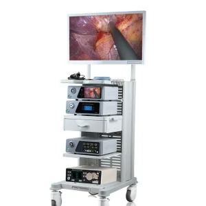
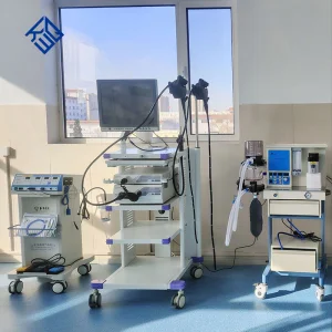
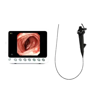
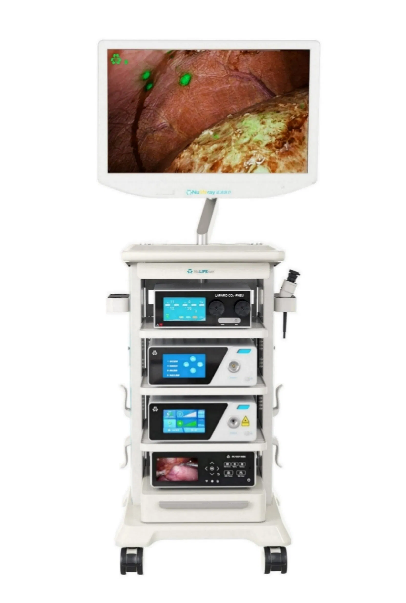
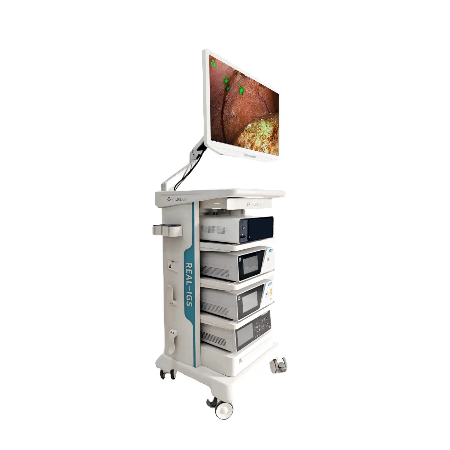
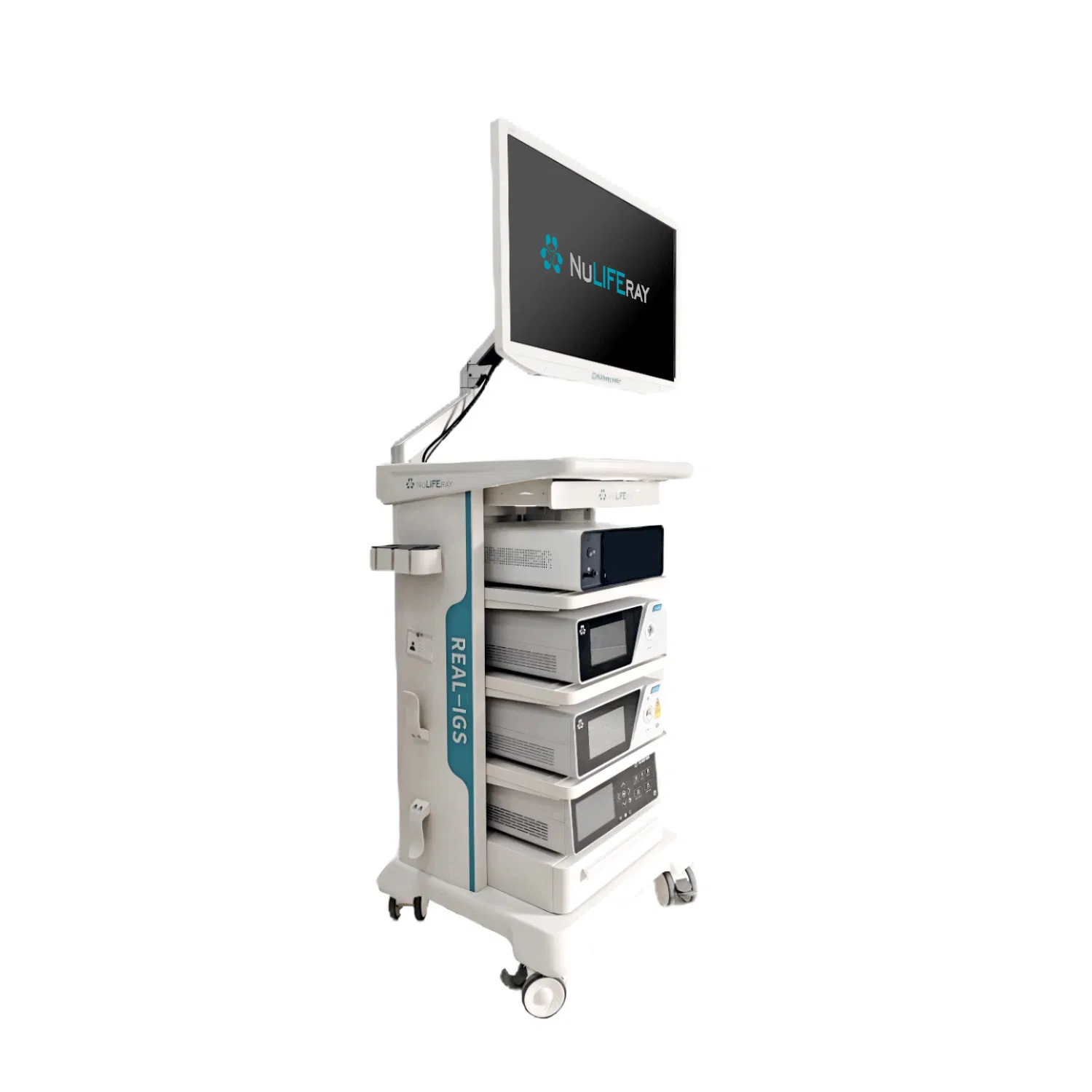
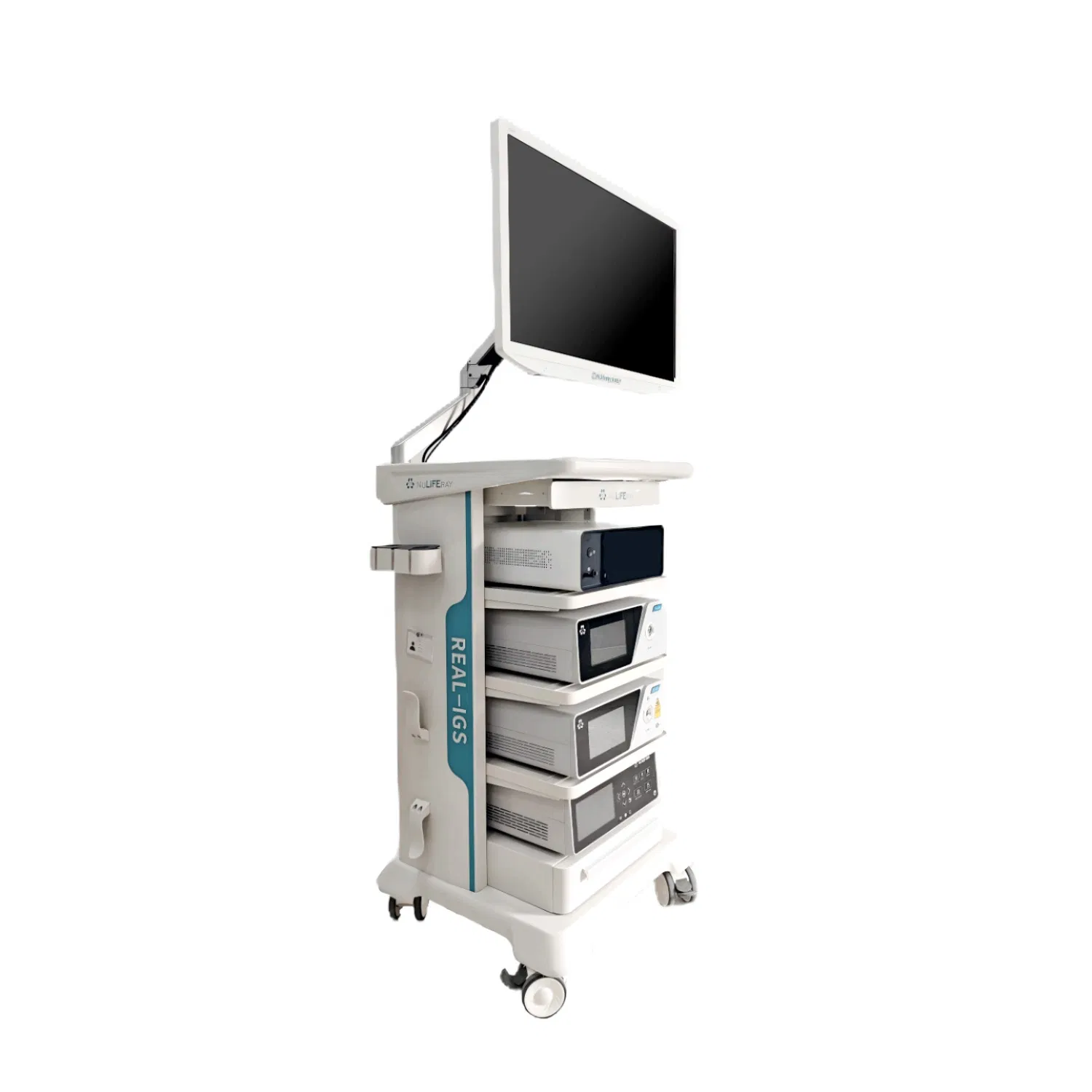
The 4K endoscopic fluorescence imaging system can not only be used as a 4K white light endoscope system to meet the clinical needs of various departments for ultra-high-definition endoscopes, but also can be used in combination with the ICG fluorescent tracer. It provides one-click selection of color images, black and white fluorescent images, color fluorescent images, and multiple images in minimally invasive endoscopic surgery. It can also switch lymphatic imaging mode, organ imaging mode, and lesion imaging mode with one click, and reserve custom modes according to the needs of the operator.
The system can also be equipped with a data workstation to achieve quantitative analysis of color gamut images and fluorescence intensity. It is suitable for dynamic observation of lesions, anatomical structures, and blood flow and lymphatic flow in minimally invasive laparoscopic surgery. Its spatial resolution allows surgeons to observe lesions, blood flow (ischemic intestinal wall judgment, anastomotic blood flow), lymphatic flow (pre-tracing sentinel lymph nodes, regional lymph nodes), and anatomical structures (liver segments, gallbladder, lung segments) in real-time.

| Parameter Item | Specifications |
|---|---|
| Laser source pulse width | N/A (continuous laser emission) |
| Chip structure | 4CMOS |
| Handle function keys | 4 remote control buttons |
| Optical adapter | Fixed focus/zoom |
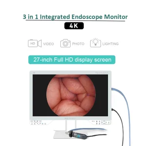
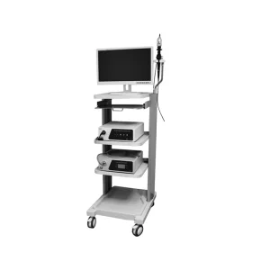
| Monitor size | 32 inches and above |
| Monitor resolution | ≥3840×2160P / 4096×2160P |
| Pixels and frame rate | ≥8.29 million pixels, 60 FPS |
| Endoscope | 0° or 30° field of view, 5mm or 10mm |
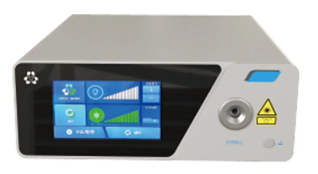
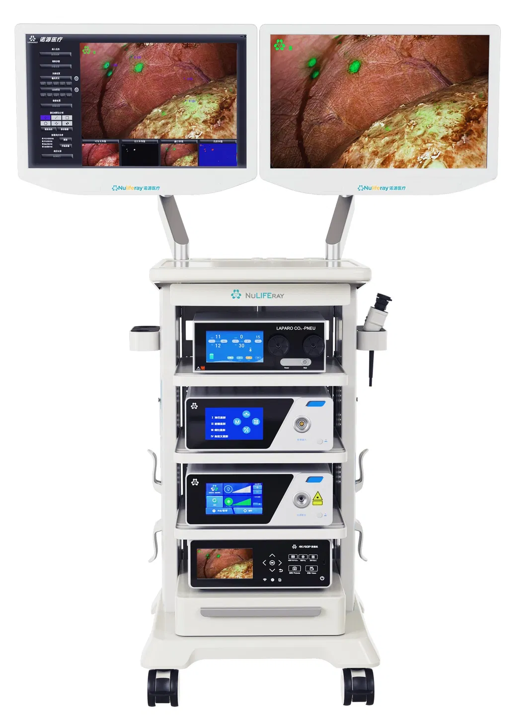
P1: Independent dual light source
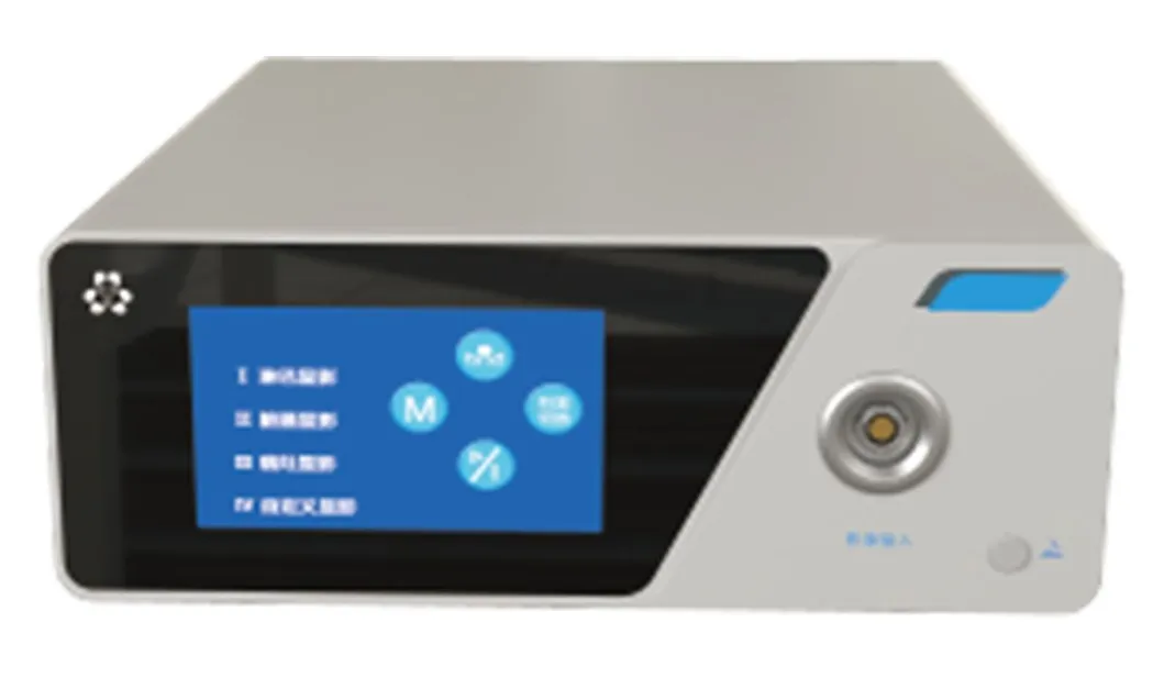
P2: Ultra HD Camera System
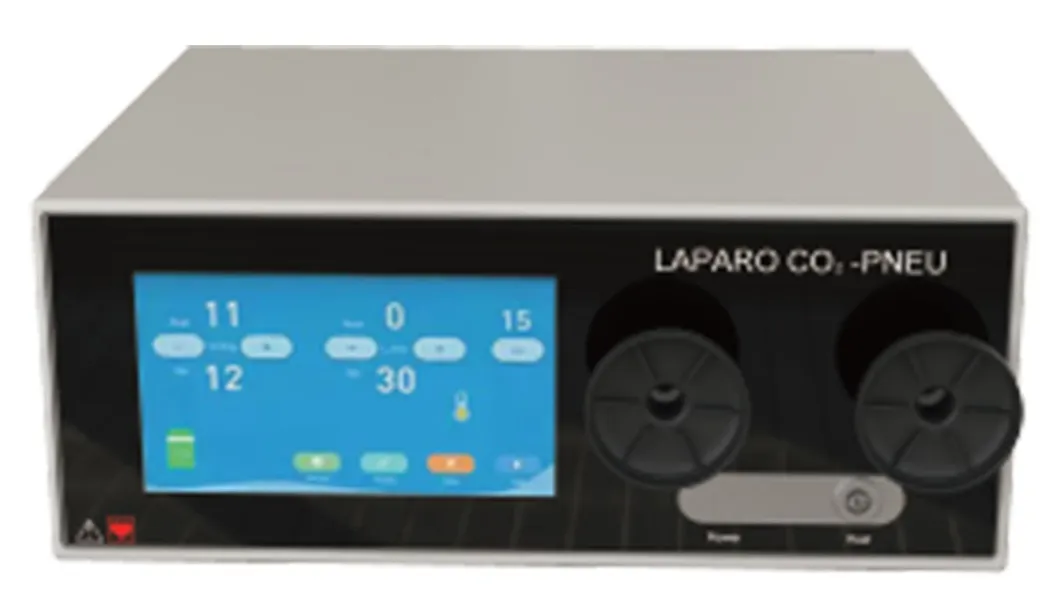
P3: Fully automatic insufflation system
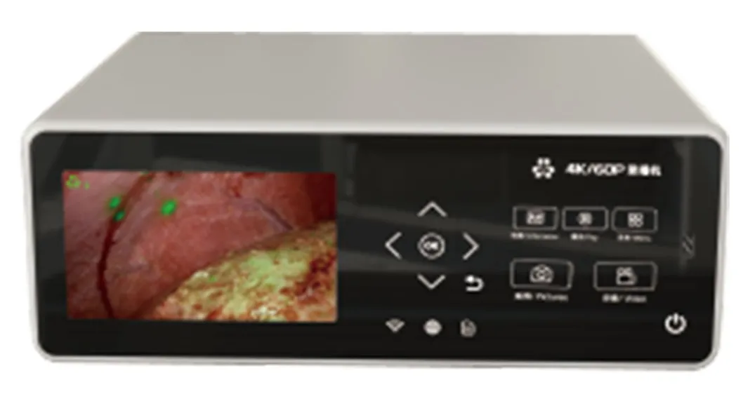
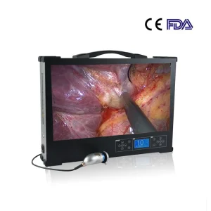
P4: True 4K recording & broadcasting
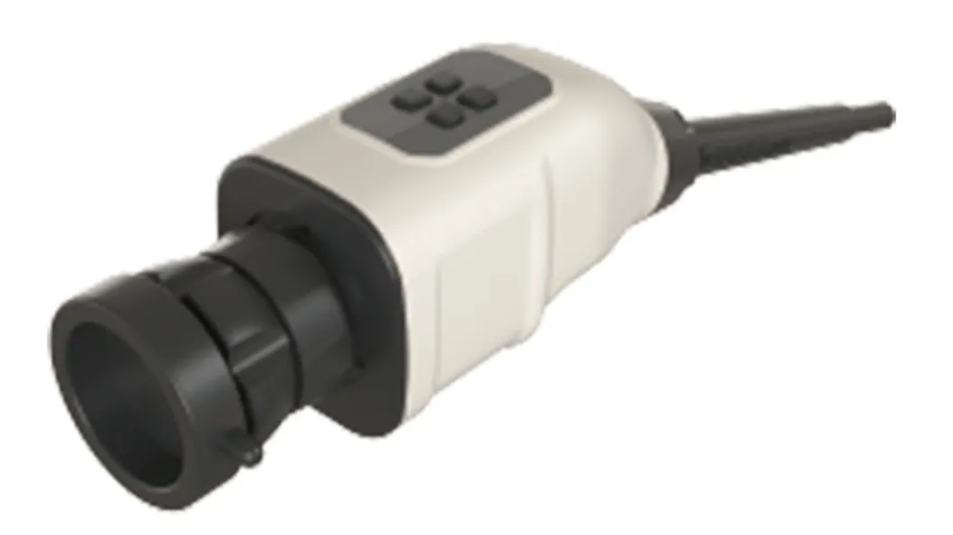
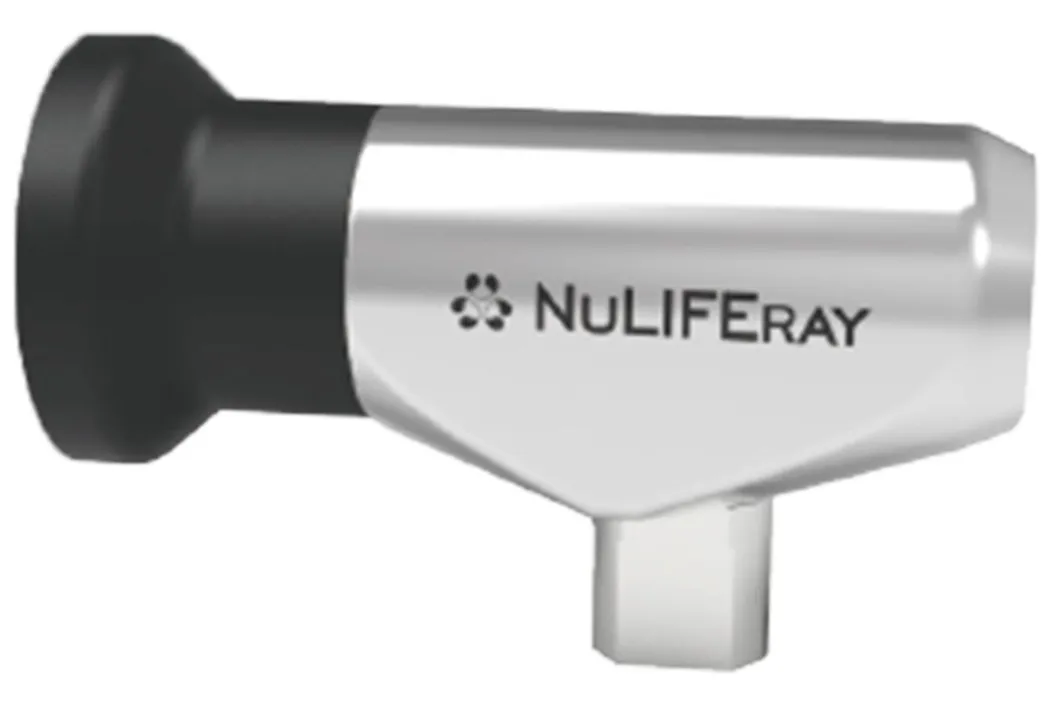
P5: 4CMOS 4K handheld camera
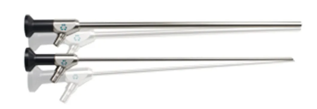
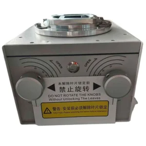
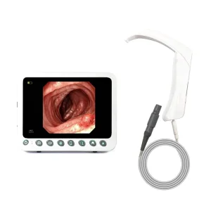
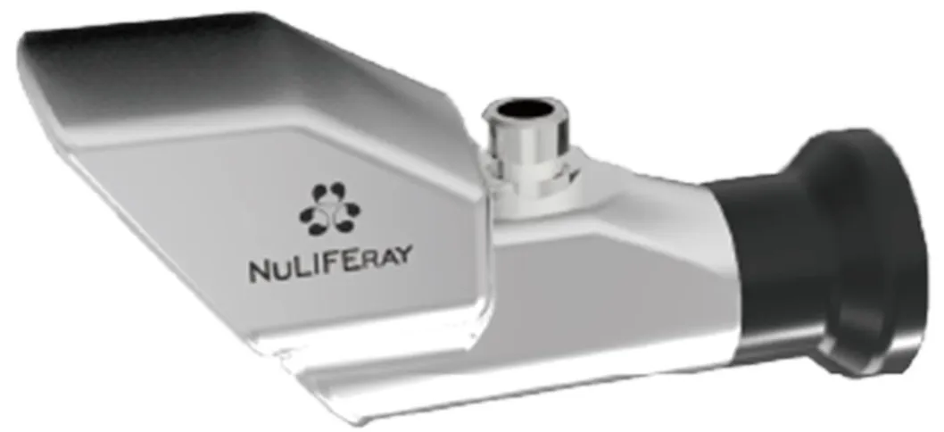
P6: Antireflective fluorescent endoscope
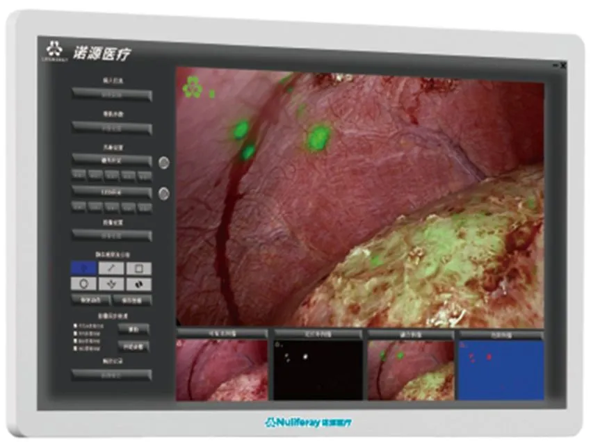
P7: Embedded software for workstation
P8: 0° Highly sensitive objective
P9: 28° Highly sensitive objective
Used in conjunction with medical endoscopes and the fluorescence dye indocyanine green (lCG) to provide illumination and real-time visible light imaging and near-infrared fluorescence imaging in minimally invasive endoscopic surgery.
Real-time liver cancer diagnosis, liver segment imaging, tumor margin evaluation, and complex biliary tract identification.
Tumor localization, sentinel lymph node labeling, and evaluation of anastomotic blood supply.
Locating lymph nodes, marking inter-segmental borders, and evaluating esophageal reconstruction blood flow.
Real-time identification of ureter and urethra, tracing pelvic tumor sentinel lymph nodes.
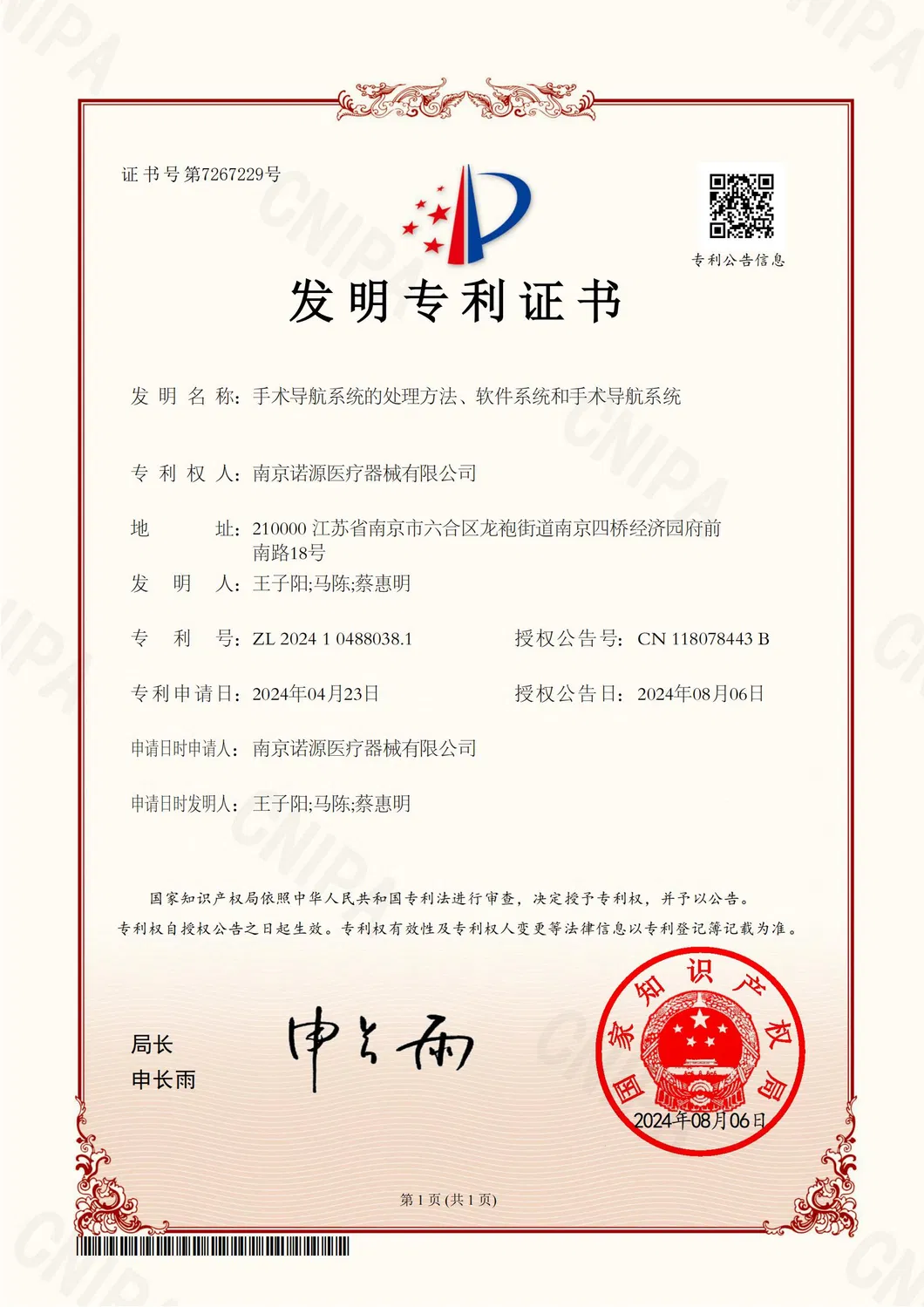


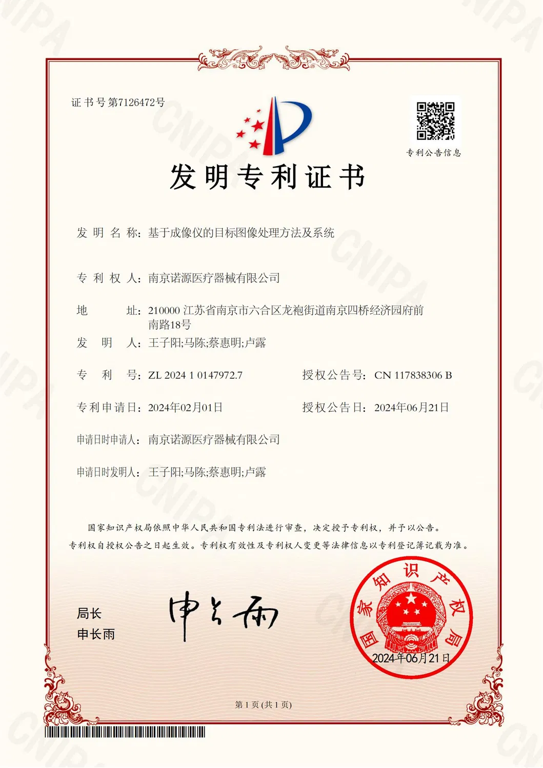
We hold 131 core invention patents and 7 US invention patents. As a "little giant" enterprise, we have undertaken numerous national and provincial R&D projects in molecular fluorescence.
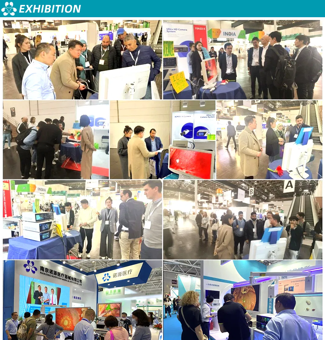

Strategic partnerships with international leading companies in Germany and Japan, serving customers in over 10 countries.